Diagram Of Chest Area - Pin on Health. In this article we will focus on: The chest workout for huge, defined pecs. Anatomy of the brain functions Diagram of a chest tube draining fluid from a plural effusion. Diagram of chest area posted on july 2, 2015 by admin when we are doing cpr why do compress the sternum area not rh quora com human chest anatomy chest diagram of the chest area including lungs, heart (hidden by the lungs) and ribcage.
ads/bitcoin1.txt
Chest pain or discomfort that is new, worsening, or occurs at rest. Experiencing such type of feeling near your chest needs to worry because it can be pointing you towards the muscular chest pain. Diagram of chest area : Thoracic cavity, also called chest cavity, the second largest hollow space of the body.it is enclosed by the ribs, the vertebral column, and the sternum, or breastbone, and is separated from the abdominal cavity (the body's largest hollow space) by a muscular and membranous partition, the diaphragm.it contains the lungs, the middle and lower airways—the tracheobronchial tree—the heart. They can give more information about injuries or diseases of the chest organs.
SAN DIEGO LYMER: Pain in areas of lymph nodes from 4.bp.blogspot.com Find out from webmd about other health problems that could be to blame. The ribs and sternum make up what is called the 'ribcage.' the ribcage protects the lungs, blood vessels, and heart. In this article we will focus on: Costochondritis, sometimes called costosternal syndrome or anterior chest wall syndrome, merely indicates pain and tenderness in the costochondral junction, which is the area along the sides of the breastbone where the ribs attach. This is an emergency situation as it can precede a heart attack, serious abnormal heart rhythm, or. Chest pain or discomfort that is new, worsening, or occurs at rest. The circulatory system does most of its. Thoracic cavity, also called chest cavity, the second largest hollow space of the body.it is enclosed by the ribs, the vertebral column, and the sternum, or breastbone, and is separated from the abdominal cavity (the body's largest hollow space) by a muscular and membranous partition, the diaphragm.it contains the lungs, the middle and lower airways—the tracheobronchial tree—the heart.
The epidermis is the outermost layer that provides a protective, waterproof seal over the body.
ads/bitcoin2.txt
Your pectoralis major and pectoralis minor muscles make up most of the muscle mass in your chest. Costochondritis, sometimes called costosternal syndrome or anterior chest wall syndrome, merely indicates pain and tenderness in the costochondral junction, which is the area along the sides of the breastbone where the ribs attach. This is an emergency situation as it can precede a heart attack, serious abnormal heart rhythm, or. Any diaphragm pain can, therefore, be very alarming. Skeletal, cardiovascular, nervous and lymphatic systems. Create your own brilliant, custom venn diagrams for free with canva's impresively easy to use online venn diagram maker. Muscle anatomy app 12 photos of the muscle anatomy app best muscle anatomy app, best muscle anatomy app. Best chest muscles diagrammatic sketch 9 photos of the best chest muscles diagrammatic sketch chest muscle diagram, chest muscle diagram exercise, female chest muscle diagram, human anatomy, chest muscle diagram, chest muscle diagram exercise, female chest muscle diagram. Sensory information from the body and critical signals. Chest pain or discomfort that is new, worsening, or occurs at rest. Thoracic cavity, also called chest cavity, the second largest hollow space of the body.it is enclosed by the ribs, the vertebral column, and the sternum, or breastbone, and is separated from the abdominal cavity (the body's largest hollow space) by a muscular and membranous partition, the diaphragm.it contains the lungs, the middle and lower airways—the tracheobronchial tree—the heart. They can give more information about injuries or diseases of the chest organs. Strains and inflammation in the chest wall muscles or ribs usually goes away on its own.
For many, the chest is made up of a single rigid bone called the sternum.however, this is not true.other than the sternum, there are other bones in the chest region, such as the ribs and even the spine at the back. Sensory information from the body and critical signals. This is an emergency situation as it can precede a heart attack, serious abnormal heart rhythm, or. In fact every radiologst should be an expert in chest film reading. And the other set of networks were.
Costochondritis - Physiopedia from www.physio-pedia.com Thoracic cavity, also called chest cavity, the second largest hollow space of the body.it is enclosed by the ribs, the vertebral column, and the sternum, or breastbone, and is separated from the abdominal cavity (the body's largest hollow space) by a muscular and membranous partition, the diaphragm.it contains the lungs, the middle and lower airways—the tracheobronchial tree—the heart. Costochondritis, sometimes called costosternal syndrome or anterior chest wall syndrome, merely indicates pain and tenderness in the costochondral junction, which is the area along the sides of the breastbone where the ribs attach. In this article we will focus on: Chest pain or discomfort that is new, worsening, or occurs at rest. Diagram of normal airway anatomy, frontal view. The chest workout for huge, defined pecs. Any diaphragm pain can, therefore, be very alarming. And the other set of networks were.
In fact every radiologst should be an expert in chest film reading.
ads/bitcoin2.txt
The chest workout for huge, defined pecs. The ribs and sternum make up what is called the 'ribcage.' the ribcage protects the lungs, blood vessels, and heart. The circulatory system does most of its. Chest pain or discomfort that is new, worsening, or occurs at rest. The sternum, or breastbone, is a flat bone at the front center of the chest. Diagram of chest area posted on july 2, 2015 by admin when we are doing cpr why do compress the sternum area not rh quora com human chest anatomy chest diagram of the chest area including lungs, heart (hidden by the lungs) and ribcage. Ct scan is a type of imaging test. Chest pain doesn't always mean you're having a heart attack. A woman's chest — like the rest of her body — is covered with skin that has two layers. Diagram of chest area posted on july 2, 2015 by admin when we are doing cpr why do compress the sternum area not rh quora com human chest anatomy chest diagram of the chest area including lungs, heart (hidden by the lungs) and ribcage. Your pectoralis major and pectoralis minor muscles make up most of the muscle mass in your chest. The epidermis is the outermost layer that provides a protective, waterproof seal over the body. Thoracic cavity, also called chest cavity, the second largest hollow space of the body.it is enclosed by the ribs, the vertebral column, and the sternum, or breastbone, and is separated from the abdominal cavity (the body's largest hollow space) by a muscular and membranous partition, the diaphragm.it contains the lungs, the middle and lower airways—the tracheobronchial tree—the heart.
Any diaphragm pain can, therefore, be very alarming. Diagram of chest area : Diagram of chest area posted on july 2, 2015 by admin when we are doing cpr why do compress the sternum area not rh quora com human chest anatomy chest diagram of the chest area including lungs, heart (hidden by the lungs) and ribcage. Location of chest pain during angina or heart attack diagram in this image, you will find an upper chest, substernal radiating to neck and jaw, substernal raiding down left arm, substernal radiating down left arm, epigastric radiating to neck, jaw, and arms, neck and jaw, left shoulder and down both arms, intrascapular in it. Best chest muscles diagrammatic sketch 9 photos of the best chest muscles diagrammatic sketch chest muscle diagram, chest muscle diagram exercise, female chest muscle diagram, human anatomy, chest muscle diagram, chest muscle diagram exercise, female chest muscle diagram.
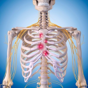
Three dimensional medical illustration of male chest ... from c8.alamy.com The nervous system of the thorax is a vital part of the nervous system as a whole, as it includes the spinal cord, peripheral nerves, and autonomic ganglia that communicate with and control many vital organs. Costochondritis, sometimes called costosternal syndrome or anterior chest wall syndrome, merely indicates pain and tenderness in the costochondral junction, which is the area along the sides of the breastbone where the ribs attach. Anatomy of the brain functions A woman's chest — like the rest of her body — is covered with skin that has two layers. The chest is the area of origin for many of the body's systems as it houses organs such as the heart, esophagus, trachea, lungs, and thoracic diaphragm. They can give more information about injuries or diseases of the chest organs. The circulatory system does most of its work inside the chest. Diagram of chest area :
Man head and chest anatomy diagram with ghost effect.
ads/bitcoin2.txt
The following diagrams describe the positions for pd & p. The circulatory system does most of its work inside the chest. The dominant muscle in the upper chest is the pectoralis major. Skeletal, cardiovascular, nervous and lymphatic systems. Thoracic cavity, also called chest cavity, the second largest hollow space of the body.it is enclosed by the ribs, the vertebral column, and the sternum, or breastbone, and is separated from the abdominal cavity (the body's largest hollow space) by a muscular and membranous partition, the diaphragm.it contains the lungs, the middle and lower airways—the tracheobronchial tree—the heart. And the other set of networks were. Focus on the target area by keeping your. For many, the chest is made up of a single rigid bone called the sternum.however, this is not true.other than the sternum, there are other bones in the chest region, such as the ribs and even the spine at the back. Chest pain or discomfort that is new, worsening, or occurs at rest. Costochondritis, sometimes called costosternal syndrome or anterior chest wall syndrome, merely indicates pain and tenderness in the costochondral junction, which is the area along the sides of the breastbone where the ribs attach. Any diaphragm pain can, therefore, be very alarming. The ribs and sternum make up what is called the 'ribcage.' the ribcage protects the lungs, blood vessels, and heart. Related posts of chest muscles diagram muscle anatomy app.